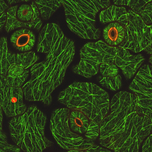
PLB Confocal Microscope
Microscope Setup:
- The LSM 710 laser scanning confocal system
- Zeiss Axio inverted microscope Observer.Z1
- Plan-Apo objectives 10x (water), 20x, 25x (water), 40x (water), and 63x (oil)
- Bright field and DIC
- X-cite wide-field fluorescent light source coupled with filter sets for blue (DAPI), green (GFP, FITC) and red (mCherry, Texas Red, Rhodamine) fluorophores
- Lasers from violet to far red: 405, 458, 488, 514, 561, 594, and 633 nm excitation wavelengths
Capabilities:
- Imaging diverse fluorophores in live or fixed samples
- Z sectioning and 3D/rotatable reconstruction
- Time-lapse recording in live samples
- Automated multi-position time-lapse
- Spectrum un-mixing (unique to Zeiss confocals)
- FRET and FRAP
Location:
- 2204 Life Sciences Building
Contact:
- Julie Lee
- 2203 Life Sciences Building
- Phone # 530-754-8139
- Email: yjlee@ucdavis.edu
Recharge
- Usage - $35 per hour
- Training - $64 per hour
